Halo® Digital Image Analysis
Transforming slide interpretation with enhanced precision and efficiency

Providing detailed, accurate, and efficient analysis of tissue samples
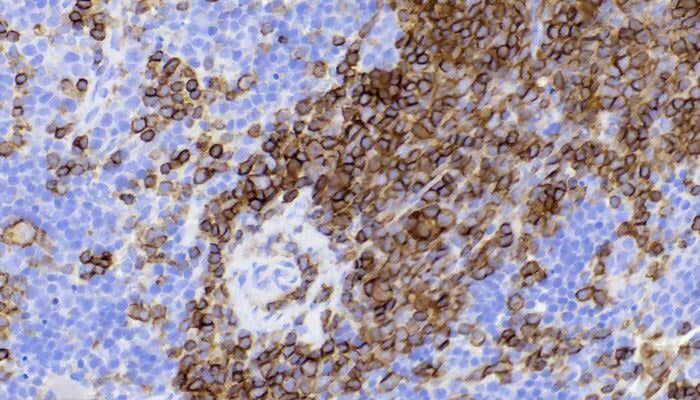
Halo Digital Image Analysis is an advanced AI-powered tool that allows for comprehensive quantification of various tissue components, such as tumor areas, necrosis, fibrosis, and cell infiltration. By accurately counting different cell types and offering spatial analysis capabilities, Halo provides detailed insights that are essential for understanding complex biological processes. Its ability to support intricate studies, including those involving CAR T-cell therapies and patient-derived xenografts (PDX), makes it an invaluable asset in advancing research and improving diagnostic accuracy.
Key benefits of Halo Digital Image Analysis
Enhanced Precision and Efficiency
Halo Digital Image Analysis significantly improves the accuracy and speed of pathology assessments, allowing for more precise measurements and faster results.
Comprehensive Quantification
The system can quantify various tissue components, such as tumor area, necrosis, fibrosis, and cell infiltration, providing detailed insights into any pathology.
Advanced Cell Counting
Combined with immunohistochemistry, Halo can accurately count different cell types, including inflammatory cells, aiding in the detailed analysis of tissue samples.
Spatial Analysis Capabilities
The Halo platform offers spatial analysis to help understand the distribution and interaction of different cell types, enhancing the overall interpretation of tissue pathology.
Support for Complex Studies
Halo is well-suited for complex studies, such as those involving CAR T-cell therapies and patient-derived xenografts (PDX), making it a versatile tool for various research applications.
Fast, accurate, and reliable results from the team you know and trust.
Connect with IDEXX BioAnalytics today.